
 1800 867 1390

As demand for skin cancer services continues to grow, many clinics are exploring collaborative...

Fulfils 50 hrs for medical professionals in Australia*
100% online
Online + workshop
Fully online: $2695
Online + workshop: from $3695
Special rates available
72.5 hours
Self-paced
2026
19 September in Sydney

General Surgeon and Lecturer, Royal Australian College of Surgeons
Associate Professor Maurice Brygel is a Melbourne general surgeon who is a pioneer in day surgery hernia repair, having performed over 10,000 hernia operations. He became known as the "hernia king" after establishing the Melbourne Hernia Clinic at Masada and Cabrini hospitals. Maurice also teaches hands-on surgical office skills, which led to him producing and presenting certificate courses in clinical procedures with HealthCert over the last five years. Maurice teaches at Notre Dame Medical School, Cabrini, the Alfred, and the Royal Australian College of Surgeons. Maurice has published extensively on hernias and authored several multimedia books including “The Video Book of Surgery” and “Exploring Essential Surgery” with the Mcgraw Hill Access Surgery and Medicine.
Senior Surgeon and Lecturer, Box Hill Hospital Melbourne
Dr Peter Grossberg is a graduate of the University of Melbourne and has a Fellowship from the Royal Australasian College of Surgeons and the American College of Surgeons. He practices as a general surgeon and an Endoscopist in Cabrini Hospital, Malvern with extensive experience in general surgery, endoscopy, and laparoscopic surgery especially laparoscopic hernia surgery. Peter has been an examiner in surgical education at the undergraduate and postgraduate level for the past 35 years. He runs workshops for surgical trainees and GPs, particularly in office procedures.
Peter has been a senior lecturer in surgery at Monash and Melbourne Universities and continues to be involved in teaching and examination at the postgraduate level for anatomy at the University of Melbourne. He has been a Senior Surgeon, Head of Unit and Chairman of the Division of Surgery at Box Hill Hospital for the past eight years.
Vascular, Endovascular & Laser Surgeon, Melbourne Vascular Surgery Unit, Cabrini Medical Centre, Victoria
Dr Peter Milne originally trained in general surgery but has specialised in vascular surgery since 1977. Peter was trained in Cardiovascular Surgery at the Methodist Hospital, Houston, Texas and in Vascular Surgery at Royal Melbourne and Epsom District Hospitals. He established the Melbourne Vascular Surgery Unit in 1985.
Peter’s expertise and special interests include aneurysm repair by open surgery and endovascular means, and carotid surgery for stroke prevention with carotid artery stenting. He also performs limb bypass surgery as well as minimally invasive techniques of angioplasty and stenting. Another speciality of Peter’s is varicose vein treatment by surgery, laser and sclerotherapy including embolisation for pelvic congestion.


As demand for skin cancer services continues to grow, many clinics are exploring collaborative...
Lip lesions are some of the trickiest skin cancers to manage. The area is small, highly visible,...
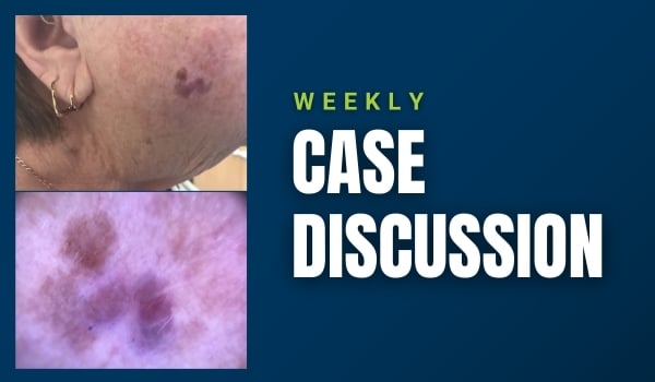
This week we revisit a case from Dr Randa Al-Hajali in which a 74-year-old female patient presents...


.png?width=100&height=100&name=Jules%20Wynyard%20edit%20circle%20(3).png "Jules Wynyard")