
 1800 867 1390
Case discussion: 5x4mm flat pigmented lesion found on posterior right leg of 65-year-old female. What do you think?
.jpg)
HealthCert Education
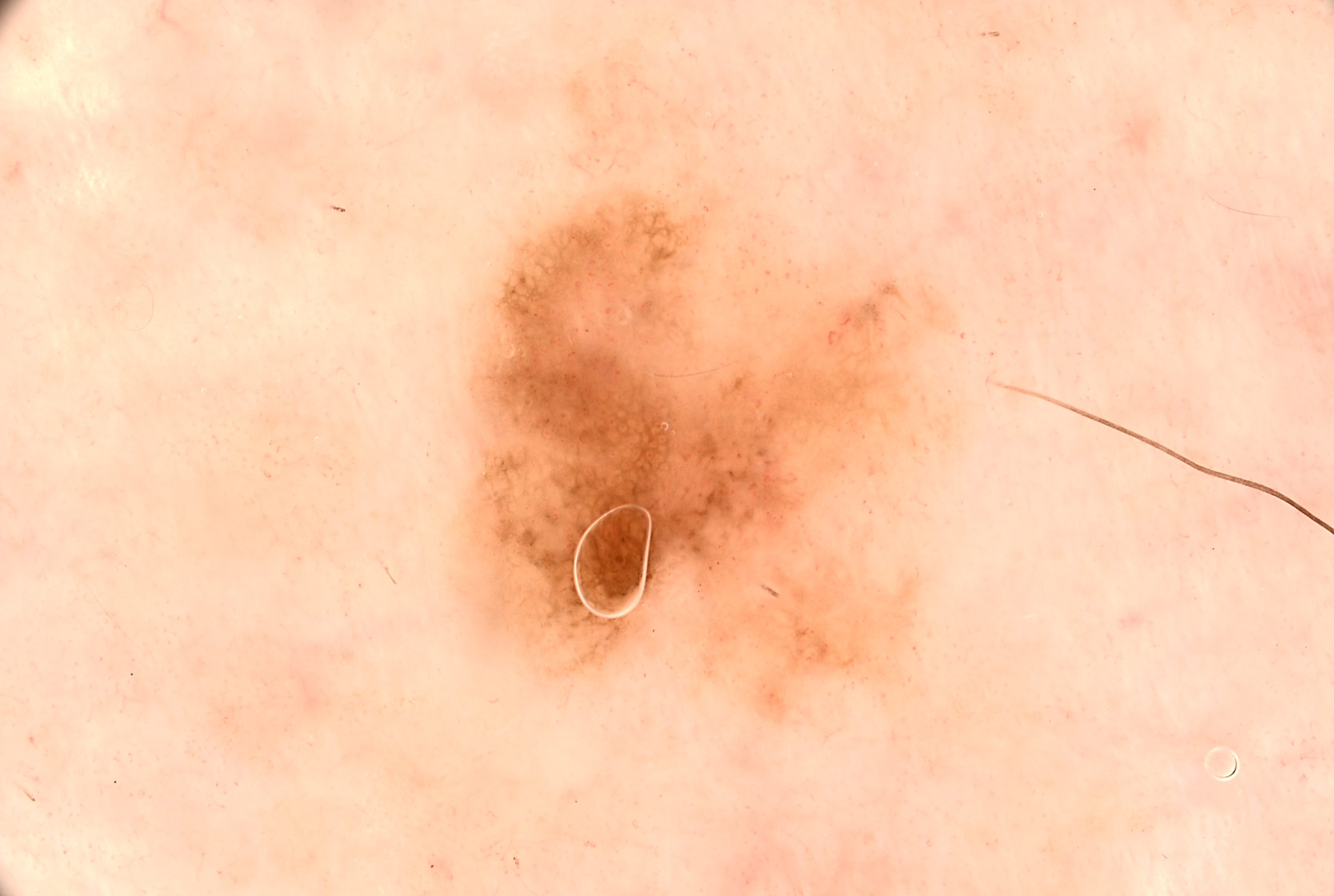
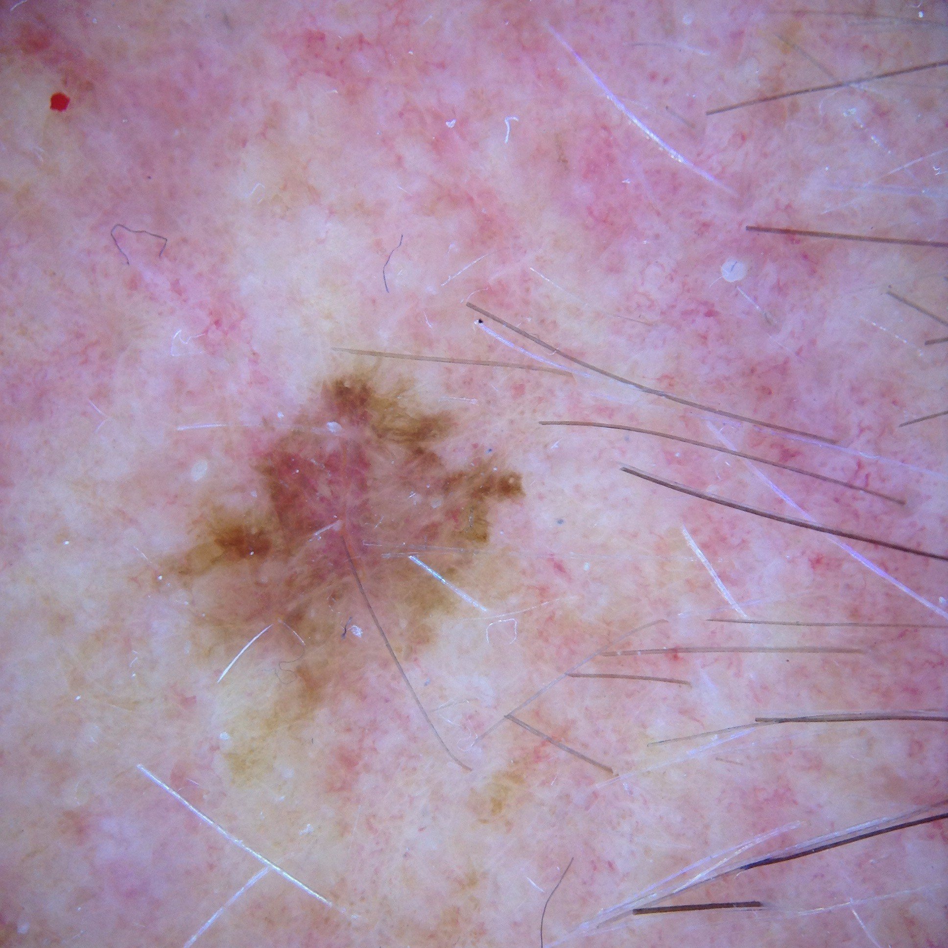
In this week's case discussion from Dr Terry Harvey, this 5x4mm flat pigmented lesion is found during a routine skin check on the posterior right leg of a 65-year-old female.
What do you think of the dermoscopy image? How do you manage this patient?
Update
An excisional biopsy with 2mm margins was performed and revealed melanoma in situ of superficial spreading subtype.
A wide local excision to achieve 5mm clinical margins was then undertaken with no complications or recurrence to date.
Interested in skin cancer medicine?
.png?width=800&height=267&name=RACGP%20newsletter%20banners%20(3).png)
 |
Participate with your cases so that we can learn together! Submit your case here or send details to admin@healthcert.com |
| Contributing to the Skin Cancer Case Discussion Blog helps meet your annual Performance Review CPD requirement! |
|
| Submit your own case* = 1 CPD hour (Performance Review) *Case must be published on the blog to qualify. |
Comment/engage with colleagues’ cases = 0.5 CPD hours (Performance Review) |
 How to claim your CPD hours How to claim your CPD hoursIf you interact with this case or submit your own case, you can Quick Log your CPD hours with the RACGP via the usual self-submission process. You will be asked to reflect on what you have learned, and you will require proof you interacted with the blog; a screenshot will suffice. |
|
.jpg)
.jpg)


.png?width=100&height=100&name=Jules%20Wynyard%20edit%20circle%20(3).png)