
 1800 867 1390
Case discussion: A 4x3mm lesion is identified on the clavicle of a 40-year-old male during a routine skin check. What do you think of it?
.jpg)
HealthCert Education
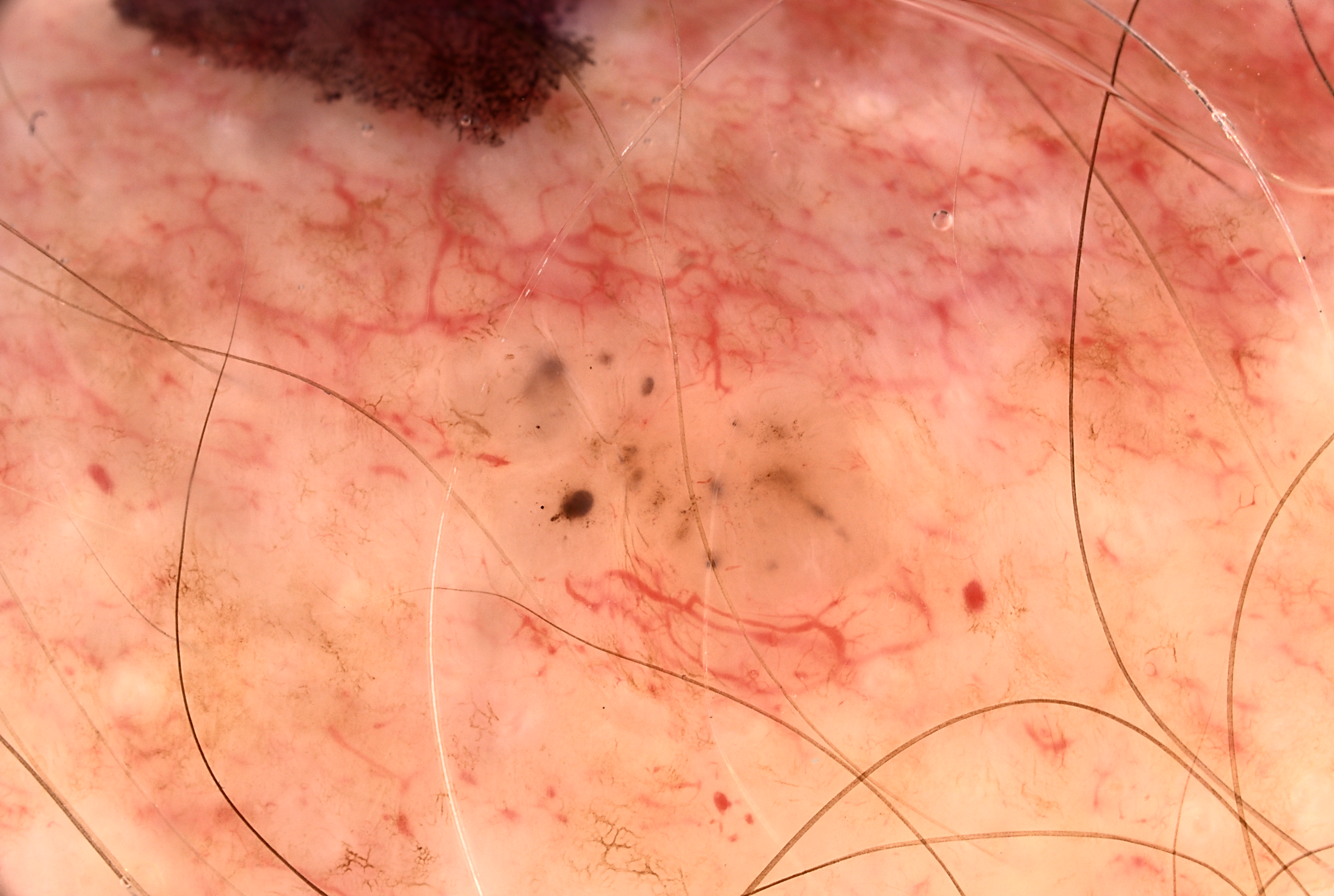
In this week's case discussion by Dr Terry Harvey, we look at a 4x3mm lesion identified on the clavicle of a 40-year-old male during a routine skin check. He was unaware of any lesion in the area.
What do you think of the dermoscopy image? What would you do next?
Update
The lesion was excised, and pathology revealed it to be a nodular and pigmented basal cell carcinoma with clear margins.
Interested in skin cancer medicine?
.png?width=800&height=267&name=RACGP%20newsletter%20banners%20(3).png)
 |
Participate with your cases so that we can learn together! Submit your case here or send details to admin@healthcert.com |
| Contributing to the Skin Cancer Case Discussion Blog helps meet your annual Performance Review CPD requirement! |
|
| Submit your own case* = 1 CPD hour (Performance Review) *Case must be published on the blog to qualify. |
Comment/engage with colleagues’ cases = 0.5 CPD hours (Performance Review) |
 How to claim your CPD hours How to claim your CPD hoursIf you interact with this case or submit your own case, you can Quick Log your CPD hours with the RACGP via the usual self-submission process. You will be asked to reflect on what you have learned, and you will require proof you interacted with the blog; a screenshot will suffice. |
|

.jpg)
.jpg)


.png?width=100&height=100&name=Jules%20Wynyard%20edit%20circle%20(3).png)