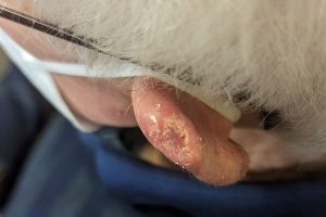
On the scalp and lips, lesions that raise concern for squamous cell carcinoma (SCC) are often not classic nodules, but flat, eroded areas with scale and vascular change. In older patients with chronically sun-damaged skin, these findings can be difficult to interpret, even with dermoscopy.
In this final instalment of a two-part series, Dr Gabriella Brancaccio focuses on the grey areas, where SCC is part of the differential but not always the diagnosis. On the scalp, SCC can closely resemble erosive pustular dermatosis of the scalp (EPDS). Both may present as superficial erosions on damaged skin, and clinically they can look almost identical.
Missed part one of this series? Catch up here.
In the video, Dr Brancaccio walks through cases where dermoscopy becomes essential, particularly when lesions appear similar at first glance. She highlights how colour cues can be more helpful than structure alone, and why biopsy is often required (sometimes more than once) before malignancy can be confidently excluded. This is especially relevant in patients with a history of scalp trauma or previous treatments.
Lesions on the lip present a different challenge. Early actinic change and inflammatory cheilitis can share features with SCC, including white areas, scale and visible vessels. Dr Brancaccio shows how these findings alone are not enough to make the diagnosis. Crusting can obscure key dermoscopic features, and true concern tends to arise when a nodular or opaque mass becomes apparent.
Watch the video to learn how to approach lesions that don’t fit neatly into a diagnostic category, with a focus on practical decision-making in general practice — using dermoscopy where it helps, recognising its limits, and knowing when uncertainty alone is enough to justify biopsy or referral.
Watch the full video
Next steps in your learning journey
🎓 Micro-Courses in Dermoscopy
Learn the essentials of skin lesion recognition in short, CPD-accredited online Micro-Courses. ➡️ Browse Micro-Courses >
🎓 Certificate Courses in Dermoscopy
Our structured pathway to elevate your skills in dermoscopy, in collaboration with the International Dermoscopy Society. ➡️ Explore full program >
🎓 HealthCert 365 subscription
Prefer flexible learning across many topics? Access 4,000+ CPD hours on-demand with HealthCert 365 — any time, any topic, one flat annual fee. ➡️ Discover HealthCert 365 >
Explore more free educational content in Dermoscopy
Dr Gabriella Brancaccio
Dr Gabriella Brancaccio is a doctor of medicine at Dermatology Unit, Università degli Studi della Campania “Luigi Vanvitelli” (University of Campania) in Naples, Italy. She is on the Executive Board of the International Dermoscopy Society as part of the Scientific Project Committee.
CPD self-submission
You can self-record CPD for this blog. Quick Log CPD hours with the RACGP/ACRRM via the usual self-submission process. You will be asked to reflect on what you have learned, and you will require supporting evidence such as a screenshot. For more information, view the: RACGP CPD guide | ACRRM CPD guide

 1800 867 1390
.jpg)
.jpg)
.jpg)


.png?width=100&height=100&name=Jules%20Wynyard%20edit%20circle%20(3).png)